Brain Function
What is the importance of brain function in the development and persistence of Alcohol Use Disorders?
The way people’s brains are wired can increase their risk for alcohol misuse and development of alcohol use disorders. Harmful alcohol use can affect the structure and function of your brain which impacts your thinking (impulse control and decision making) and behavior.
The key takeaway: Some individuals from families with many members affected with alcohol use disorder may show differences in brain functioning before they ever drink, and are at higher risk for problematic drinking and developing alcohol use disorder. In addition, chronic and hazardous use of alcohol has harmful effects on the brain and its functioning, which impacts thinking and behavior.
Brain structure and function are involved in the development and persistence of alcohol use disorders
Changes in the structure and functioning of the brain have long been known to accompany chronic and harmful alcohol use. Some of these brain changes are known to recover as a person remains sober, while others persist and lead to addiction. These brain differences involve impairments in specific brain networks, or areas of the brain that work together closely. Networks that are involved in the ability to control an impulse and make decisions, respond to reward, and regulate emotion have been shown to be impacted in alcohol use disorder. Different brain networks are involved in the different stages of alcohol use disorder. These brain changes affect reward/emotion regulation and decision making which contribute to the development and persistence of alcohol use disorders. [See this report for details: THE NEUROBIOLOGY OF SUBSTANCE USE, MISUSE, AND ADDICTION].
The way people’s brains are wired can increase their risk to develop alcohol use disorders
Some of the same differences in brain function seen in those with alcohol use disorder are also seen in some of their children, before they ever drink any alcohol, especially in families with many members affected by alcohol use disorder (AUD). These children are more likely to show differences in the same brain networks that impact decision making, reward, and emotion as those adults involved in increased drinking behaviors and alcohol use disorder, and their brains may mature more slowly. These differences in brain function, which tend to travel in families, put these individuals at higher risk for problematic drinking and the development of alcohol use disorder.
Brain waves and neurocognitive tests provide sensitive measures of brain function and patterns of development involved in AUD and increased risk
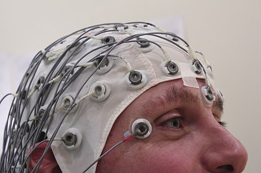 The brain is always active, even during sleep, as the brain cells, or “neurons” are constantly in communication with each other in networks, causing electrical fluctuations in the brain called brain waves. These brain waves or “neural oscillations” are detected using a method called the (sidenote: EEG is a method to record the electrical activity of the brain using electrodes placed along the scalp.) The brain’s natural EEG can be recorded while people are resting. The brain’s electrical activity can also be measured while an individual is performing a simple task, such as pressing a button when they see a specific picture shown on a computer screen while ignoring other pictures. Brain waves recorded during a task or event are called event-related potentials (ERPs) and event-related oscillations (EROs). ERP and ERO analysis allow us to evaluate different ways the brain works, such as identifying specific images or sounds, decision making, or emotional processing. Some studies of brain waves at rest and during tasks show how different areas of the brain communicate and work together, like musicians in an orchestra; when brain areas are not working together, a person’s thinking and behavior may suffer.
Studying brain waves helps us to detect and understand the impact of heavy drinking on brain functioning, such as decision making, the ability to control an impulse, how people respond to rewards, and memory, as well as existing differences in these brain functions in those at higher risk for AUD before they ever drank alcohol. These brain wave measures, examined along with other risk and protective factors for AUD, and the effects of alcohol consumption on the development of the adolescent human brain, can be used to identify and understand the factors that may put an adolescent at greater risk for an alcohol use disorder.
Brain Function, AUD risk, neurodevelopmental trajectories and neurogenomic studies in COGA
Early studies of young sons of fathers with AUD found that they had similar brain wave features as their fathers and other adults with AUD, even before they drank alcohol; later studies showed similar findings in daughters and sons of both mothers and fathers with AUD. These studies focused on an ERP brain wave, called the (sidenote: It is usually the largest brain wave component of the event-related potentials (ERP), which typically occurs around/after 300 ms and reflects processing of the task’s context and/or demand, including stimulus evaluation and decision making.) a large positive wave that reaches a peak shortly after performing a mental task, such as pressing a button to only certain images while ignoring others (Begleiter et al 1984). This finding showed that brain wave abnormalities observed in children of fathers with AUD may be a marker of higher risk for developing AUD, and not simply a result of years of heavy drinking. The idea that some brain wave measures may indicate higher risk that was passed along from parent to child changed how we study AUD and was a main motivation to launch the COGA project. From its inception 30 years ago, a primary emphasis of the COGA project has been to study brain function as a measure of higher risk in families with alcohol use disorder (AUD) including their relatives and children. We have also studied genetic and environmental influences on these brain function measures and how they affect a person’s risk of AUD or their resistance to it throughout adolescent and young adult development and continuing throughout the lifespan.
What are some of the major findings associated with brain waves and AUD from the COGA study?
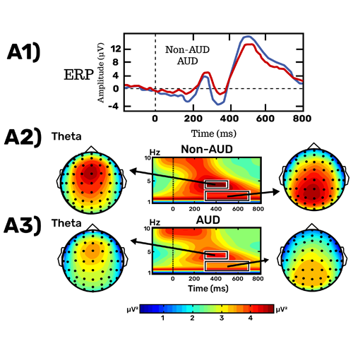
- A relatively smaller P300, a specific part of the ERP brainwave, while pressing a button only to a target images was found in those with AUD (Jones et al., 2006) and their younger high-risk relatives (Rangaswamy et al., 2007). A weaker signal in slow brain waves called delta (1-3 Hz or cycles per second) and theta (4-7 Hz) during the same task was also found in both those with AUD and high-risk relatives (Jones et al., 2006; Rangaswamy et al., 2007).
- Compared to people without AUD, individuals with long-term AUD showed reduced brain activity at the frontal region while inhibiting or trying to stop a button press in a Go/NoGo task (Pandey et al., 2012).
- Another COGA study found that childhood sexual trauma negatively impacted how brain waves changed during inhibitory control with development, which in turn, may increase risk for AUD and related mental health problems (Meyers et al., 2019a).
- Excessive fast brain waves, called beta frequency band (13-29 Hz), was found in both adults with AUD (Rangaswamy et al., 2002) and their high-risk offspring (Rangaswamy et al., 2004).
- Abnormal brain network communication has also been found in those with alcohol use disorder (Kamarajan et al., 2020).
- Using machine learning algorithms, several EEG measures were identified as risk factors for the development of AUD (Kinreich et al. 2021) and other EEG measures were related to remission from AUD (Kinreich et al. 2021).
What are some of the major findings with brain waves and genetic factors from the COGA study?
- Significant linkage for EEG fast brain waves and GABRA2 on chromosome 4 also associated with AUD and related disorders was found (Porjesz et al., 2002; Edenberg et al., 2004).
- Significant linkage and association with GABRA2 and interhemispheric theta coherence, which refers to synchronized brain activity between brain regions in slow waves (3-7 Hz) was also reported (Porjesz and Rangaswamy, 2007; Rangaswamy and Porjesz, 2008).
- We also reported significant linkage between the theta and delta (1-3 Hz) oscillations underlying P300 to target images and CHRM2 on chromosome 7, which we found is also associated with AUD and related disorders (Porjesz and Rangaswamy, 2007; Rangaswamy and Porjesz, 2008).
- A genome-wide association study (GWAS) of brain functioning measured by theta wave activity during a target-detection task identified the role of genetic variants in KCNJ6 gene on chromosome 21 (Kang et al., 2012).
- Another GWAS study by COGA identified the role of genetic variants on chromosome 3 in regulating fast beta waves and alcohol use behavior (Meyers et al., 2017).
- A recent GWAS study identified genetic variants on chromosome 18 that were involved in regulating neural communication (theta wave connectivity) and alcohol use behavior, potentially via dysregulated myelination (Meyers et al., 2020).
- Using polygenic risk scores (PRS), genetic risk factors in the cholinergic system were found to influence brain development of brain wave patterns and neural communication during adolescence and young adulthood (Meyers et al., 2019b).
